Microsurgery & Complex Reconstruction
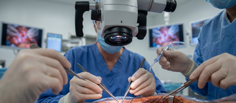
Microsurgery is a highly advanced subspecialty of reconstructive surgery that involves repairing and connecting microscopic structures—such as blood vessels and nerves measuring less than a few millimeters in diameter—using specialized operating microscopes, ultra-fine needles, and hair-thin sutures. When trauma, cancer removal, congenital anomalies, or chronic infections leave behind extensive tissue voids or complex wounds that cannot be closed by standard means, microsurgery allows for the transfer of healthy tissue from one part of the body to another to restore both form and vital function.
The hallmark of complex reconstruction is tissue engineering and restoration. Rather than simply covering a wound, microsurgery transfers living, vascularized composites of skin, fat, muscle, or bone, seamlessly reconnecting the tissue’s blood supply at the new site to ensure its permanent survival.
By removing excess, hanging skin and targeted fat deposits, an arm lift tightens the structural tissue framework from the underarm area to the elbow, restoring a toned, sculpted, and youthfully proportioned arm profile.
Quick Facts
Surgery Time
4 to 8+ hours depending on the anatomical complexity and the number of microvascular connections required.
Anesthesia
General anesthesia, continuously managed to maintain optimal blood pressure and tissue perfusion.
Stay Required
3 to 7 nights in a specialized inpatient or intensive care setting for continuous, hourly monitoring of tissue vascularity.
Initial Recovery
2 to 3 weeks for primary wound healing; 6 to 12 weeks for transferred muscles or bones to integrate structurally and regain functional strength.
Major Microsurgical Procedures & Clinical Applications
Microsurgical techniques are deployed across various anatomical regions to solve complex reconstructive challenges:
- Microvascular Free Tissue Transfer (Free Flaps)
Unlike local flaps that are swung from adjacent skin, a “free flap” involves completely detaching a composite block of tissue from a distant donor site and transplanting it to the recipient site by reconnecting its individual artery and veins:
- DIEP Flap (Deep Inferior Epigastric Perforator): Transfers skin and fat from the lower abdomen to reconstruct a natural, soft breast after a mastectomy for breast cancer, leaving the abdominal core muscles completely intact.
- Fibula Free Flap: Replaces segments of the mandible (jawbone) or long bones of the limbs following tumor resection or severe trauma, using the vascularized fibula bone from the lower leg.
- Radial Forearm Free Flap: Utilizes thin, pliable skin and a segment of the radial artery from the forearm to reconstruct delicate linings inside the oral cavity, tongue, or pharynx.
- Anterolateral Thigh (ALT) Flap: Provides robust soft tissue and skin coverage from the outer thigh to reconstruct extensive defects on the scalp, face, extremities, or trunk.
- Peripheral Nerve Repair & Reconstruction
- Digital Nerve Repair: Reconnecting severed nerves in the fingers following sharp lacerations to restore fine sensation.
- Brachial Plexus & Nerve Transfers: Re-routing healthy, redundant nerves or utilizing nerve grafts (such as the sural nerve) to bypass severely damaged nerve networks in the arm or leg, restoring lost muscle function and movement.
- Replantation & Revascularization
- Digit and Limb Replantation: The immediate surgical reattachment of completely severed fingers, hands, or limbs following traumatic accidents, meticulously setting bones, repairing tendons, and using microvascular suturing to immediately restore blood circulation.
The Precision Approach to Microvascular Surgery
Step 1: Radical Debridement & Preparation
The recipient site is completely cleared of any damaged tissue, infection, or tumor cells, and the local recipient artery and veins are carefully isolated under magnification.
Step 2: Flap Harvesting
The distant donor tissue is meticulously dissected, protecting its single feeding vascular pedicle (the main artery and veins) until the recipient site is entirely prepared.
Step 3: The Micro-Anastomosis
Using ultra-fine 9-0 or 10-0 sutures (thinner than a human hair), the surgeon sews the donor artery and veins to the recipient vessels, establishing an immediate, living blood supply.
Step 4: Functional Inset & Closure
The transferred tissue is precisely shaped and secured into the defect space, ensuring there is zero kinking or tension on the newly repaired blood vessels.
The Post-Operative Monitoring & Recovery Timeline
- The Profile: This is the most critical phase of microsurgical recovery. The transferred tissue is completely dependent on the newly sewn micro-vessels.
- Hourly Monitoring: The nursing and surgical teams perform hourly assessments of the flap, checking its temperature, color, capillary refill time, and utilizing specialized acoustic Doppler probes to listen to the arterial and venous blood flow.
Positioning: You will remain resting in a temperature-controlled room with the reconstructed area supported and immobilized to prevent any pulling or shifting of the microvascular pedicle.
- Clinic Visit: Surface skin sutures from both the reconstruction site and the donor site are evaluated and comfortably removed between days 10 and 14.
- Gentle Mobilization: Progressive, non-weight-bearing movement is introduced based on the location of the surgery. If a muscle or bone was transferred, tailored physical therapy begins during this window to preserve joint flexibility.
- Tissue Adaptation: Over several months, the transferred tissue loses its early post-op swelling and naturally contours to its new environment. Nerve regeneration across the flap edges occurs slowly, with protective sensation gradually returning over 6 to 12 months.
Side Effects vs. Warning Signs
|
Expected Normal Symptoms
|
Warning Signs (Call the Clinic Immediately)
|
|---|---|
|
Expected widespread swelling, bruising, and local tissue firmness
|
The transferred tissue suddenly changes color, turning pale white, dusky blue, or dark purple
|
|
Temporary numbness across the flap surface and donor site lines
|
The flap feels notably cold to the touch compared to neighboring skin. A fever rising above 101°F (38.3°C)
|
|
Light pink or serous fluid tracking lightly onto surgical dressings
|
A sudden, massive swelling or firm, painful bulge forming beneath the incisions
• Loss of the audible Doppler pulse or a sudden change in tissue firmness
|
Frequently Asked Questions (FAQ's)
Trusted guidance to help you feel informed and confident about your surgical journey..
- This is known as flap compromise and most commonly occurs within the first 48 hours, which is why monitoring is so intensive. If the hourly checks show a change in tissue color or blood flow, the patient is immediately returned to the operating room. The surgeon opens the connection, clears any microscopic clots, and re-does the microvascular suture lines to save the tissue. When caught early, these interventions are highly successful.
- Yes, absolutely. Plastic surgeons select donor sites very carefully, choosing areas that have redundant tissue, muscle, or bone function. For example, when the fibula bone is harvested from the leg, the upper and lower joints of the ankle and knee are left entirely intact, allowing you to walk, run, and bear weight completely normally after initial physical rehabilitation.
- A skin graft is a very thin shaving of skin that has no independent blood vessels; it relies entirely on absorbing nutrients from the bed of the wound it is placed upon, making it suitable only for shallow wounds. A free tissue transfer is a deep, three-dimensional block of living tissue (incorporating skin, fat, muscle, or bone) that carries its own functional artery and veins, allowing it to bridge massive, deep structural voids where bone or tendon is exposed.
- While hospital discharge typically occurs within 5 to 7 days once the flap is deemed stable, full recovery depends on the structures involved. For soft tissue reconstructions (like a DIEP flap breast), most patients return to normal daily routines within 6 weeks. For bony reconstructions (like a fibula jaw repair), full functional recovery and return to heavy physical activity can take 3 to 6 months, supported by continuous physical therapy.